
Breast Cancer Screening
What are the Guidelines for Breast Cancer Screening?
ACS recommendations for breast cancer screening-
- Women age 40 and older should have a mammogram every year
- Women in their 20s and 30s should have a clinical breast exam (CBE) preferably every 3 years. Starting at age 40, women should have a CBE every year.
- Breast self-exam (BSE) is an option for women starting in their 20s.
- Women who are at high risk for breast cancer should get an MRI and a mammogram every year.
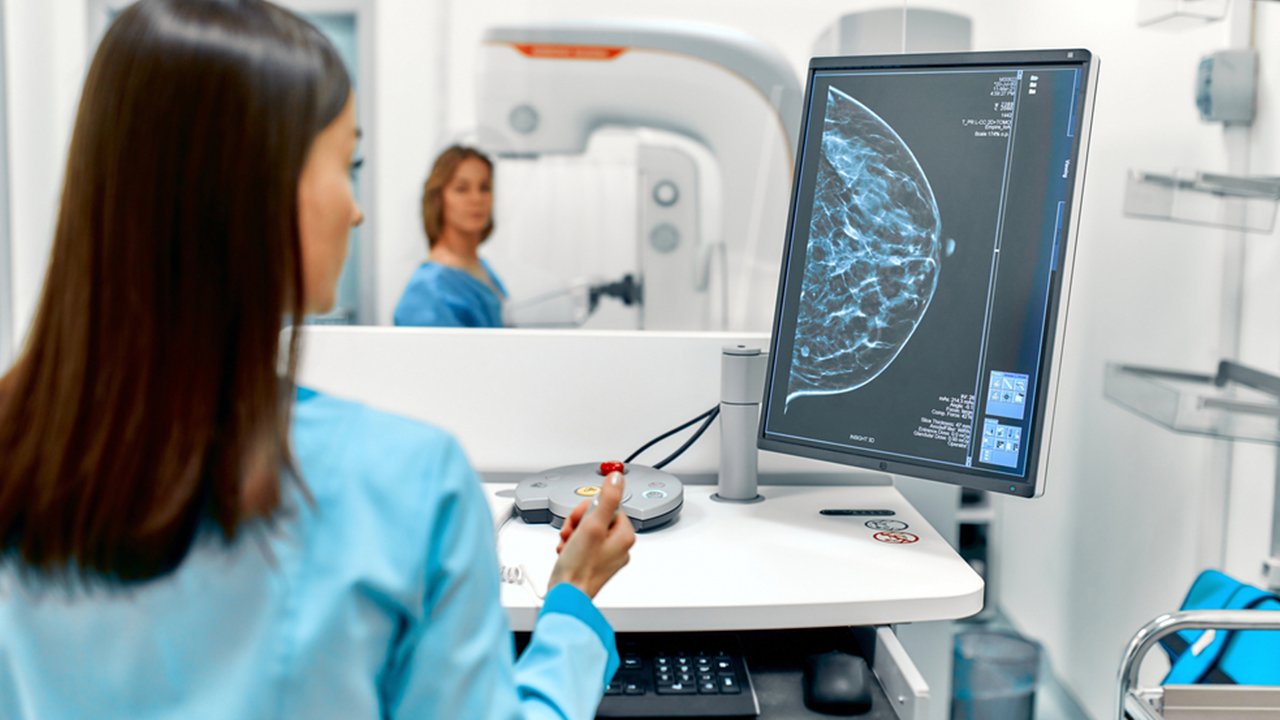
What is the role of Mammography in Breast Cancer Screening?
Mammography is being used for screening since 1980’s. In 1990s, a decrease in breast cancer mortality of 2.1% was reported by the American Cancer Society. In the 15 years from 1983 to 1998, the rate of discovery of stage I breast cancer more than doubled and that of in situ carcinoma more than tripled while the incidence of stage IV disease at diagnosis fell by almost one half.
It has two views, Craniocaudal (CC) and Mediolateral Oblique (MLO) to characterize the type and location of breast lump. Breast compression is done between the plates which increases image contrast, decreases radiation dose, reduces motion, and minimizes superimposition of tissues. Pain and discomfort may be addressed by soft radiolucent breast cushion, premedication with oral analgesics and topical lignocaine
It can detect cancer 1.5-4.0 years before it is clinically evident. Mean glandular dose is 0.1-0.2 rads (1-2 mGy) per exposure, and effective rose received is around the same as that from normal background radiation for 3 months.
Film screen vs Digital Mammography
According to Digital Mammographic Imaging Screening Trial (DMIST) and Oslo II study, overall diagnostic accuracy was similar but digital was more accurate for premenopausal, perimenopausal and women with dense breasts.
Digital mammography, when available, may offer a small screening advantage in women younger than 50 years old.
Abnormalities on mammography
- The most specific mammographic feature of malignancy is a spiculated focal mass. Positive Predictive Value of mass with a spiculated margin is 81% and with irregular shape is 73%.
- The density of a non-calcified mass. 70% of masses with high density were malignant, and 22% with low density were malignant.
- Clustered microcalcifications are seen in approximately 60% of cancers detected mammographically.
- Linear branching microcalcifications have a higher predictive value for malignancy than do granular microcalcifications, especially for high grade DCIS.
- However, breast cancers, including DCIS, more often present with the granular type of calcifications.
Breast density
All reports have a statement regarding the breast density. Most radiologists use the four categories described in the BI-RADS atlas, based on the proportion of glandular (radiodense) tissue with respect to fatty (radiolucent) tissue. The four main categories are:
- Predominantly fatty (0 to 25 percent dense)
- Scattered fibroglandular densities (25 to 50 percent dense)
- Heterogeneously dense (51 to 75 percent dense), and
- Dense (greater than 75 percent)
What are new techniques for Breast Cancer Screening?
Positron Emission Mammography (PEM) – It has a sensitivity 86 to 91% and specificity 91 to 93%. PEM does not reliably detect low grade malignancies
Breast Specific Gamma Imaging (BSGI)– It uses gamma cameras with 2 to 3 mm in-plane resolution in a mammographic configuration to provide images of the breast. It is based on the observation that breast cancers accumulate technetium-99m sestamibi in intracellular mitochondria. BSGI demonstrated equal sensitivity and greater specificity than MRI for the detection of breast cancer
Breast Tomosynthesis – Breast tomosynthesis (“3-D mammography”) has been approved by the US Food and Drug Administration for routine clinical use. It is a modification of digital mammography and uses a moving X-ray source and digital detector. A three dimensional volume of data is acquired and reconstructed using computer algorithms to generate thin sections of images.
In the screening setting, this helps to decrease recall rates by delineating true lesions from spurious lesions caused by overlapping structures seen on routine mammography. In the diagnostic setting, tomosynthesis improves lesion characterization, thereby decreasing the need for biopsy, leading to fewer false positive biopsies and higher rates of cancer detection.
You Can also read: Exercises To Do After Breast Cancer Surgery
What is the role of Clinical Breast Examination in Screening?
The sensitivity of CBE is considerably less than mammography average of 54%. The American Cancer Society states that its continuing recommendation to perform CBE is based on lack of conclusive evidence against it and the opportunity to use the examination as a time to discuss early breast cancer detection and other breast cancer issues.
Recommendations For CBE
- The American Cancer Society recommends clinical breast examination every three years from age 20 to 39, and annually thereafter
- The American College of Obstetricians and Gynecologists recommends clinical breast examination every one to three years from age 20 to 39, and annually thereafter
- The US Preventive Services Task Force concludes that evidence is insufficient to assess additional benefits of clinical breast examination beyond mammography
- The World Health Organization does not recommend clinical breast examination
You Can Also Read: Risk Factors For Breast Cancer
What is the role of MRI Breast in Screening?
American Cancer Society Indications For Breast MRI-
- Known BRCA mutation carriers
- First degree relatives of known BRCA mutation carriers
- Women with an approximate lifetime risk of breast cancer from 20 to over 25 percent
- Had radiation therapy to the chest when they were between the ages of 10 and 30 years
- Have Li-Fraumeni syndrome, Cowden syndrome, or Bannayan-Riley-Ruvalcaba syndrome, or have first-degree relatives with one of these syndromes.